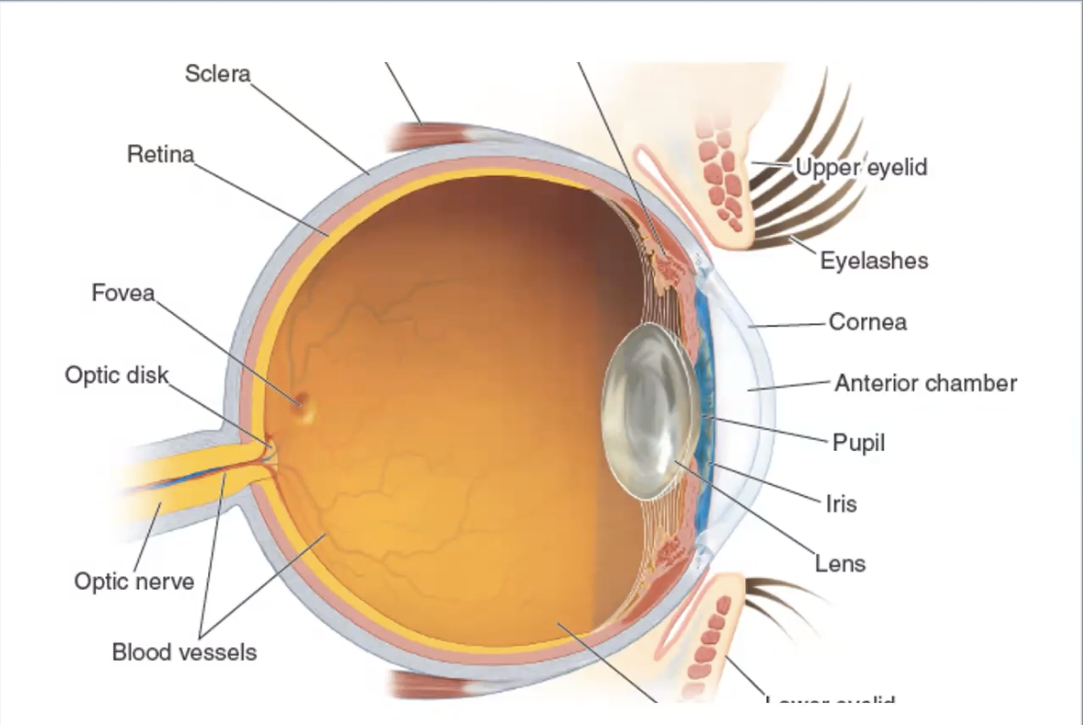
Lens focuses light on the retina in back of eye.
Cornea begins process of bending light.
Retina features fovea, wherel ight is focused when lookingat detailed image
optic disk, axons on retinal cells leave, no photo receptors here, each eye has tiny blind spot
Dont see it unless looking through one eye only.
- Macula imp for seeing HD images, fovea important part of it
- Macula degeneration is major cause of blindness
- blood vescles infront of retina
- red eye results from light reflecting from blood supply infront of retina
- Retina means Fishermans Net in latin
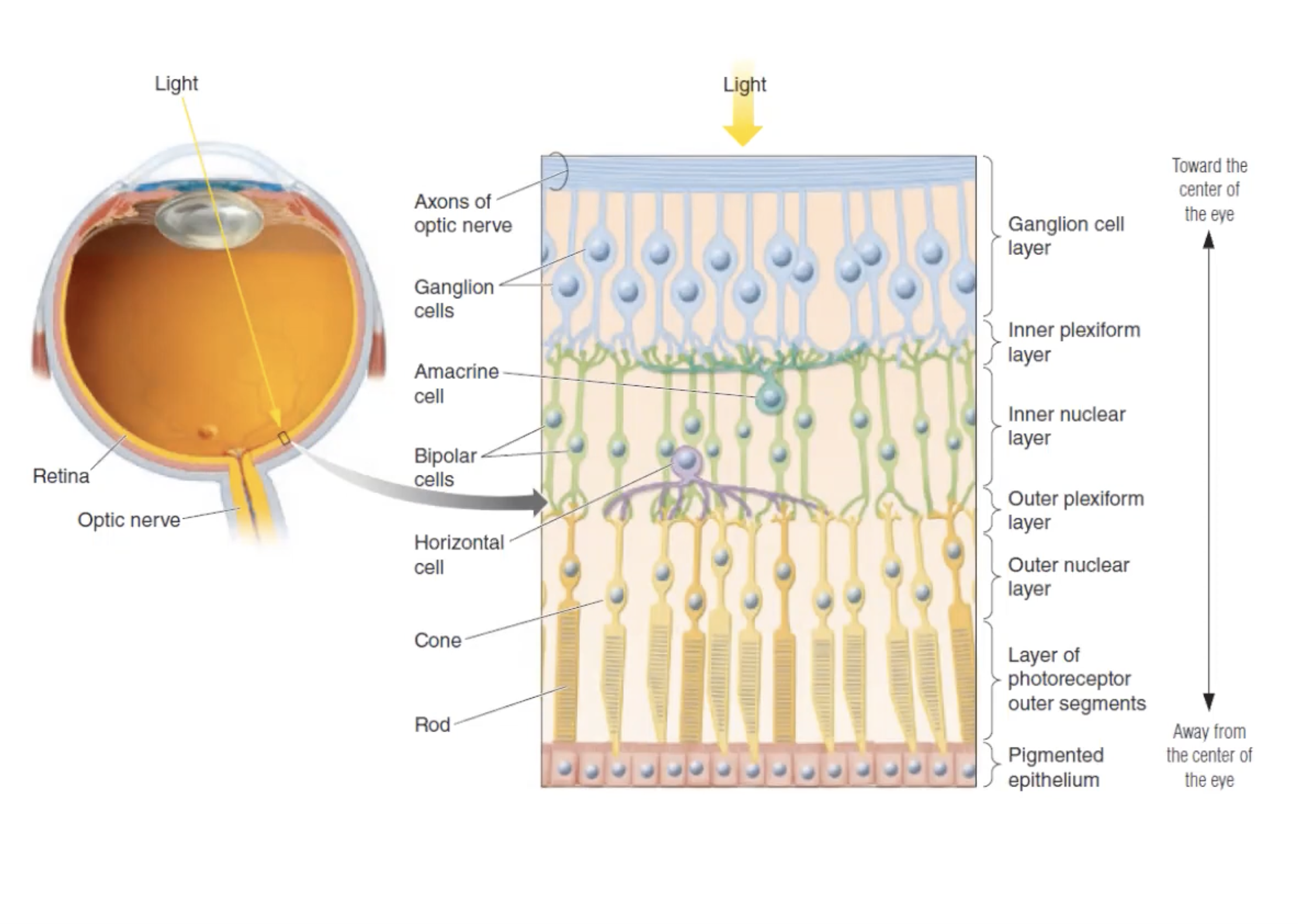
- Don't see neural layers of blood vesles cus they dont moove
- Only cells that produce action potentials are Ganglion cells
- Others produce graded potentials, vvarying in size and duration
Rods and Cones
Named after physical shape
- Rods have pointy end
- Excel at movement in dim light, not fine detail or color
- See a single photon of lige, seeing candle flame 30,000 away dark night
- cones have stacks of pancakes
- require bright light,
- color and detail
- 3 types, red, green, or blue
- Rods dominate outer eye, cones dominate inner by the macula
- more sensitive to dimlight looking off to the side
The Dark Current
- How photo receptorsthe respond to a packet of light energy
- more accurate in the dark than light
- photo receptor releaes more glutamate in dark, reduces in light
- cells know what to do with the info
Visual Receptive Fields
- Part of visual feel where light activates cell
- outside, cell is blind
- cell inside responds to light corresponding to
- each cell respnd to specific side
- Visual fields overlap have different sizes,
- Large field struggles to comment on finely detailed images
- Purpose?
- Identify borders and edges
What They Do
- Awake animal stares at screen
- moves dot on screen until cell becomes active
- this fines visual field
- cells have 3 settings: base line, increased, and decreased.
- Bipolar cell quiet in baseline, ganglion shows base line noise
- Cell isnt interested between light on and light off, focused on difference of life within its visual field
- Lateral Inhibition
- Jobs of cells is to identify edges
Visual Pathways
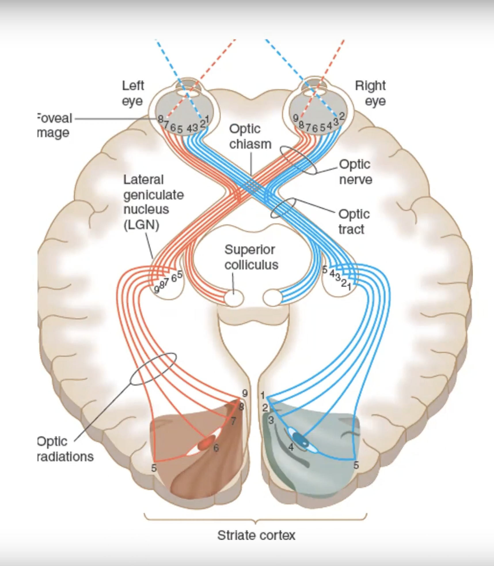
Adaptation for foreward facing eyes, encormous advantages in depth perception
- In Humans, 90% of Ganglion go to the LGN, a few go to hypothalumus, rest go to superior colliculus
Lateral Geniculate Nucleus (LGN)
Light keeos seperate from both eyes for depth perception
- Inpit from rod and cone sysyem also kept seperate
Cortical Receptive Fields
Cortical visual neurons no longer donut shaped, but have them
- respond to bars of life
- more complicated images
- cell silent on inhibitory surround
- Corticol cells snesitive to orientation
- Complex have broader perceptive field and respoind to movement in particular direction
Cortical Modules
IHubel and Wiesel's "ice cube"model of visual cortec
- Each chunk integrates nput from reeach eye
- process 180 degree of bars orientation
- Cytochrome oxidase Blobs do colour
Beyond the Cortex
Very visual animals
- Dorsal stream -- Where receed towards parietal, cordinate visual with space n movement (insert credit card)
- Ventral stream. -- What, FFA facial recognition